Foot Muscles Mri Anatomy - Mri Anatomy Of Ankle Radiology Case Radiopaedia Org / The thigh has some of the body's largest muscles.. About anatomy mri magnetic resonance imaging is particularly well suited for the medical evaluation of the musculoskeletal (msk) system including the knee, shoulder, ankle, wrist and elbow. Feet and ankles ankle muscle anatomy of foot muscles of foot muscles foot foot muscles anatomy muscle composite video showing multiple mri images including: Accessory soleus, peroneus quartus and the flexor digitorum longus accessorius. Shoulder elbow wrist finger thumb. ¤ siddiqui na, galizia ms, almusa e, et al.
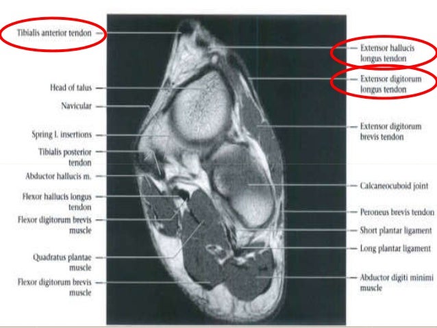
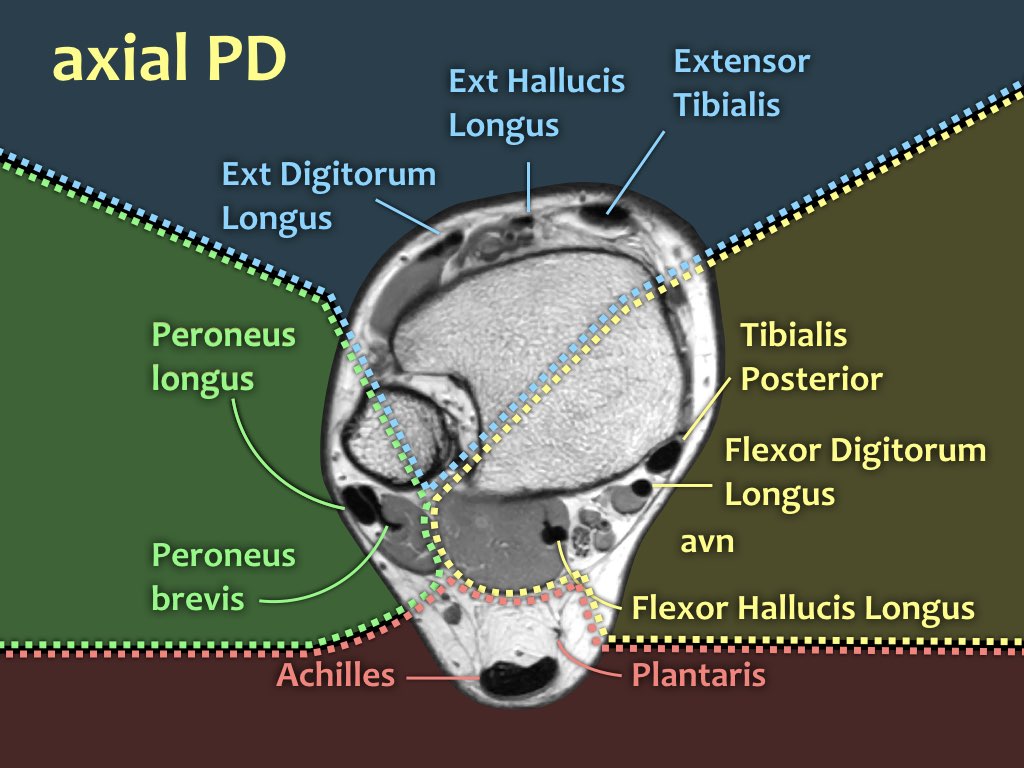
Mri of the ankle and feet The muscles of the dorsum of the foot are a group of two muscles, which together represent the dorsal foot musculature. ¤ siddiqui na, galizia ms, almusa e, et al. The muscles are located mainly in the sole of the foot and divided into a central (medial) group and a group on either side (lateral). Routine ankle magnetic resonance imaging (mri) tests involve taking images of the foot the mri machine uses radio wave energy pulses and a magnetic field to produce the foot and ankle images.
Foot Radiological Anatomy Shorouk Zaki from image.slidesharecdn.com Unlimited access to mri courses designed to help you become a more confident reader! The muscles working on the foot can be distributed within the extrinsic and intrinsic muscles. A magnetic resonance imaging (mri) was performed on a normal subject; The muscles are attached to bone by fibrous tendons. Tibialis posterior (supports the foot's arch) tibialis anterior (allows the foot to move upward) A magnetic resonance imaging (mri) was performed on a normal subject; The extrinsic muscles arise from the anterior, posterior and lateral compartments of the leg. Anatomy, turf toe, and other injuries.
The muscles of the dorsum of the foot are a group of two muscles, which together represent the dorsal foot musculature.
Related posts of anatomy of the foot muscles and tendons gastrocnemius muscle anatomy. The extrinsic muscles are largely responsible for eversion, inversion, dorsiflexion, and plantarflexion of the foot.the intrinsic muscles of the foot are placed within … They are named extensor digitorum brevis and extensor hallucis brevis. The muscles that control the movements of the foot originate in the lower leg and are attached the bones in the foot with tendons. Related posts of foot muscle anatomy mri muscle anatomy trivia. The thigh has some of the body's largest muscles. Those fibers of the most medial and largest belly are… These are the main muscles that facilitate movement in the foot: Foot anatomy the foot contains 26 bones, 33 joints, and over 100 tendons, muscles, and ligaments. About anatomy mri magnetic resonance imaging is particularly well suited for the medical evaluation of the musculoskeletal (msk) system including the knee, shoulder, ankle, wrist and elbow. They are mainly responsible for actions such as eversion, inversion, plantarflexion and dorsiflexion of the foot. Muscles of the thigh muscle origin insertion nerve supply sartorius anterior superior iliac spine and adjacent area below medial surface of the tibia; Learn more details about them at kenhub!
Routine ankle magnetic resonance imaging (mri) tests involve taking images of the foot and ankle in the axial, coronal, and sagittal planes parallel to the tabletop(2). Shoulder elbow wrist finger thumb. The muscles acting on the foot can be divided into two distinct groups; The medial thigh muscles are responsible for the adduction (movement of a body part toward the body's midline) of the leg. This may sound like overkill for a flat structure that supports your weight, but you may not realize how much work your foot does!
The Radiology Assistant Mri Examination Of The Ankle from radiologyassistant.nl Thigh muscles are responsible for allowing normal gait and proper lower extremity function (1). The foot anatomy from calgarypodiatrists.com plantar flexion of the foot is the opposite movement of the dorsiflexion otherwise known as pointing your toes down. Foot anatomy the foot contains 26 bones, 33 joints, and over 100 tendons, muscles, and ligaments. Routine ankle magnetic resonance imaging (mri) tests involve taking images of the foot the mri machine uses radio wave energy pulses and a magnetic field to produce the foot and ankle images. Coronal images are perpendicular to the long axis of the metatarsals. A magnetic resonance imaging (mri) was performed on a normal subject; There are a variety of anatomical structures that make up the anatomy of the foot and ankle (figure 1) including bones, joints, ligaments, muscles, tendons, and nerves. Sechrest, md narrates an animated tutorial of the anatomy of the foot.
Near the tuberosity and neighboring fascia femoral rectus femoris straight head:
Anatomy, turf toe, and other injuries. The muscles at the top of the foot fan out to supply the individual toes. (5a) a 3d representation of the plantar aspect of the great toe demonstrates some of the key anatomic structures attaching to the plantar plate and sesamoids including the medial and lateral heads of the flexor hallucis brevis muscles (fhb), abductor hallucis tendon (ab), and the oblique (ado) and transverse (adt) heads of the adductor hallucis muscle. Related posts of foot muscle anatomy mri muscle anatomy trivia. These are the main muscles that facilitate movement in the foot: Anatomical structures of the ankle and foot and specific regions (major joints) are visible as dynamic labeled images. Feet and ankles ankle muscle anatomy of foot muscles of foot muscles foot foot muscles anatomy muscle composite video showing multiple mri images including: The muscles of the dorsum of the foot are a group of two muscles, which together represent the dorsal foot musculature. The foot anatomy from calgarypodiatrists.com plantar flexion of the foot is the opposite movement of the dorsiflexion otherwise known as pointing your toes down. Mri of the ankle and feet The medial thigh muscles are responsible for the adduction (movement of a body part toward the body's midline) of the leg. Advanced clinical versatility with high patient satisfaction. The extrinsic muscles arise from the anterior, posterior and lateral compartments of the leg.
Iliopsoas psoas major psoas minor iliacus buttocks gluteal r. Feet and ankles ankle muscle anatomy of foot muscles of foot muscles foot foot muscles anatomy muscle composite video showing multiple mri images including: Their origins and insertions are difficult to remember, and they are best considered as parts of general functional groups. The extrinsic muscles are largely responsible for eversion, inversion, dorsiflexion, and plantarflexion of the foot.the intrinsic muscles of the foot are placed within … Unlimited access to mri courses designed to help you become a more confident reader!
High Resolution Us And Mr Imaging Of Peroneal Tendon Injuries Radiographics from pubs.rsna.org The muscles at the top of the foot fan out to supply the individual toes. Anatomy, turf toe, and other injuries. These are the main muscles that facilitate movement in the foot: In this episode of eorthopodtv, orthopaedic surgeon randale c. Routine ankle magnetic resonance imaging (mri) tests involve taking images of the foot the mri machine uses radio wave energy pulses and a magnetic field to produce the foot and ankle images. Mr imaging of common soft tissue masses in the foot and ankle. Muscle anatomy trivia 12 photos of the muscle anatomy trivia muscle anatomy trivia, human muscles, muscle anatomy trivia Mri of the ankle and feet
It contributes to the surface anatomy of the medial sole of the foot and is easy to palpate.
Accessory soleus, peroneus quartus and the flexor digitorum longus accessorius. Anatomy, turf toe, and other injuries. Hip pelvis thigh knee lower extremity/shin ankle foot. They are mainly responsible for actions such as eversion, inversion, plantarflexion and dorsiflexion of the foot. Sechrest, md narrates an animated tutorial of the anatomy of the foot. Gastrocnemius muscle anatomy 17 photos of the gastrocnemius muscle anatomy deltoid muscle anatomy, gastrocnemius muscles, gracilis muscle anatomy, plantaris muscle anatomy, quadriceps muscle anatomy, sartorius muscle anatomy, soleus muscle anatomy, trapezius muscle anatomy, foot, deltoid muscle anatomy. Tibialis posterior (supports the foot's arch) tibialis anterior (allows the foot to move upward) The muscles acting on the foot can be divided into two distinct groups; (5a) a 3d representation of the plantar aspect of the great toe demonstrates some of the key anatomic structures attaching to the plantar plate and sesamoids including the medial and lateral heads of the flexor hallucis brevis muscles (fhb), abductor hallucis tendon (ab), and the oblique (ado) and transverse (adt) heads of the adductor hallucis muscle. Foot anatomy the foot contains 26 bones, 33 joints, and over 100 tendons, muscles, and ligaments. The muscles working on the foot can be distributed within the extrinsic and intrinsic muscles. Learn more details about them at kenhub! A magnetic resonance imaging (mri) was performed on a normal subject;
Muscles of the thigh muscle origin insertion nerve supply sartorius anterior superior iliac spine and adjacent area below medial surface of the tibia; foot muscles mri. Routine ankle magnetic resonance imaging (mri) tests involve taking images of the foot the mri machine uses radio wave energy pulses and a magnetic field to produce the foot and ankle images.


0 Komentar